Before the invention of imaging technology, doctors had no way to see what was going on underneath their patients’ skin without making an incision. Today, we have a variety of diagnostic imaging tools that allow us to see views of the body that are invisible to the naked eye, long before there’s any need to use anesthesia or pick up a scalpel.

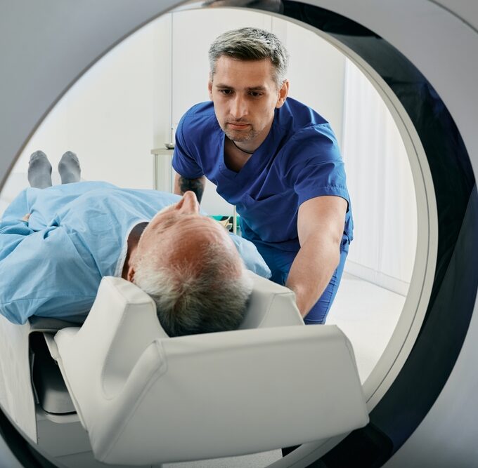
Computed tomography scans (CT) are one of the most common types of diagnostic imaging tests. At the Orthopaedic Institute of Ohio, we use CT scans to visualize the internal structures of the body, including the bones, muscles, blood vessels, tendons, ligaments and cartilage. The resulting images help guide our treatment plans and procedures and give us critical information about how well a treatment is working.

What Is a CT Scan?

CT scans, sometimes known as computed axial tomography (​CAT) scans, use sophisticated x-ray technology to produce pictures of the inside of the body. Multiple images are taken from many different angles with computer processing technology to create a “cross-sectional” view of the scanned area.

A popular way to visualize what this means is to picture a loaf of bread. The loaf can be looked at as a whole, or each slice of bread can be viewed individually. You can change how you look at the bread depending on what information you need. With CT scans, we divide the body into thin “slices” or cross-sections, and computer software reassembles them into a detailed view of the body’s interior. They may also be combined to create a 3D image of a specific area of the body. 

What Is a CT Scan With Contrast?

In some cases, we use a dye called contrast material to help internal structures show up better on CT scan images. The contrast material blocks x-rays and looks white on images, which helps to highlight structures like blood vessels in the area we’re examining.

Contrast comes in several forms. A doctor can administer it via injection or enema, or it can be consumed orally in a liquid. The method is chosen based on which area of the body is being inspected. Not all CT scans are done with contrast.

What Are Some Common Uses for CT Scans?

CT scans are noninvasive, painless and accurate. With this technology, doctors can:

  • Study internal structures in various parts of the body 
  • Diagnose musculoskeletal injuries and disorders
  • Locate masses and tumors, including cancer
  • Assess the severity of internal bleeding and injuries
  • Guide surgical procedures
  • Monitor the effectiveness of certain treatments 

What Is the Difference Between a CT Scan and an X-Ray?

In a conventional x-ray, the x-ray source stays in a fixed position. During a CT scan, the patient lies still while the x-ray source rotates around them. The scan can image many different types of tissue and structures all at the same time. CT images show greater detail and can be reformatted to create a 3D picture. This provides much more information than traditional x-rays. 

Together, these things mean CT scans give us a more complete perspective on your condition and may reveal areas of concern that do not show up on other tests. 

What Is the Difference Between a CT Scan and an MRI?

Unlike CT, which uses radiation, MRI (magnetic resonance imaging) uses radio waves and a magnet to create detailed images of the interior of the body. MRI excels at showing certain conditions that CT scans cannot detect. The procedure is longer and noisier than a CT scan, which some patients do not tolerate well. MRI cannot be performed on people with certain implanted medical devices.

CT scans are shorter and less sensitive to patient movement than MRI. CT is safe even if you have an implanted medical device. Though a CT scan does deliver radiation, the amount is small, and the potential benefits of the test typically outweigh the potential risks from radiation.

How Should I Prepare for a CT Scan?

As a general rule, preparation is minimal before a CT scan. Wear comfortable, loose-fitting clothing to your exam. It is best not to wear items with metal pieces such as zippers and studs. You will need to remove any metal objects, like jewelry and hairpins, from your body before the scan. 

Depending on the part of your body to be scanned, you may be asked not to eat or drink for a few hours before your appointment. Inform your doctor if you are taking any medications, as some medications should be stopped before a CT scan. Let your doctor know about any recent illnesses or medical conditions and your medical history. Women should inform their doctor if they are pregnant or if there is any possibility that they may be pregnant.

What Can I Expect During and After a CT Scan?

A CT scanner looks like a large doughnut standing upright. You will rest on a table that moves through the opening of the “doughnut” into a tunnel. Straps, pillows and/or a head cradle may be used to help you stay in position. 

While the table maneuvers you into the scanner, an X-ray tube will rotate around you. You may hear buzzing and whirring noises, but you will not feel anything. A technologist will conduct the scan from a separate room. They will be able to see you, and you will be able to communicate with them via intercom. 

Your CT scan may take only a few minutes. After the appointment, you will be free to return to your normal routine. You may receive special aftercare instructions if you were given contrast material for the scan. If you had a sedative, someone will need to accompany you home.

Are CT Scan Results Immediate?

CT images are stored as electronic files and reviewed by a radiologist. The radiologist analyzes the images and sends a report to the doctor who ordered the exam. If the radiologist detects any abnormalities, you may need additional tests or treatments. The results of a CT scan usually take 24 hours. 

CT scans are a powerful diagnostic tool that helps our providers accurately diagnose your health concerns and provide the right treatment for you. For more information about OIO’s diagnostic testing services or to schedule an exam, contact the Orthopaedic Institute of Ohio at 419-222-6622.